Your chance to ask a young researcher about his work
Arman Eshaghi will answer your questions
Last updated: 3rd December 2015
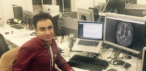
Arman Eshaghi, a researcher from Iran, was awarded one of MSIF’s McDonald Fellowships in 2014. This award enables talented young MS researchers from emerging countries to work with leading researchers in MS.
Arman is currently based in UCL Institute of Neurology in London, working with Prof Olga Ciccarelli, Prof Alan Thompson, and Prof Daniel Alexander. He will answer questions on his area of expertise (Predicting disease progression using MRI and computer models) on MSIF’s Facebook account on Wednesday 16 December.
Please send your questions for Arman to support@msif.org. Keep in mind that the Question & Answer session will be about Arman’s research, and not clinical advice. You will also be able to ask questions via Facebook. To make sure you receive all the information on how to take part, please sign up to Act on MS, using the form at the right side of this page (bottom of the page if you are on a mobile device).
Here is a little more information on Arman’s research, in his own words:
“Grouping patients into different MS subtypes (e.g. relapsing-remitting or progressive) is important when selecting appropriate treatments. Currently the decision is based on clinical evaluation, which is subjective and unreliable. MRI, on the other hand, provides an objective assessment of brain, but it isn’t clear whether we can use MRI to predict progression or group patients into subtypes.
My research (a collaboration between the Institute of Neurology and UCL Department of Computer Science) aims to study MRI in detail to describe MS progression. I will specifically look at brain tissue changes over time to develop an objective way to describe the stage of a person’s MS.
I will combine advanced image analysis and statistical models to explore brain tissue loss in each region to predict people who are at higher risk of disability later in their disease course. It will provide a unique way to look into each MS subtype based on a set of characteristics shown on MRI. By combining information from imaging, other biomarkers (such as spinal fluids) and clinical information we could identify people who are at risk of higher disability earlier and assign the correct treatment.”